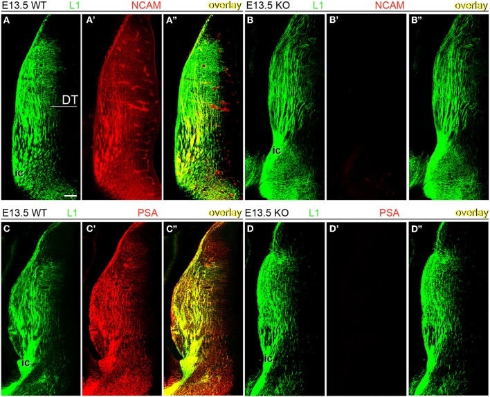
Figure 2.
Expression of NCAM and oligopolysialylated NCAM in thalamocortical axons in the dorsal thalamus at E13.5. (A–A”) TC axons co-labeled by L1 and NCAM antibodies in wild type mice. (B–B”) In NCAM null mutant mice, NCAM immunostaining was absent, confirming the specificity of monoclonal antibody P61 in our material. (C–C”,D–D”) PSA immunoreactivity was present in L1-immunoreactive axons in the dorsal thalamus of wild type mice (C) but absent in NCAM null mice (D), suggesting that PSA is linked to NCAM in these axons. L1-immunoreactive TC axons formed descending fascicles that converged into the caudal limb of the internal capsule (ic) leading through the ventral telencephalon to the developing cortex. Single confocal optical sections. DT, dorsal thalamus; ic, internal capsule. Bar (in A): 100 μm.

