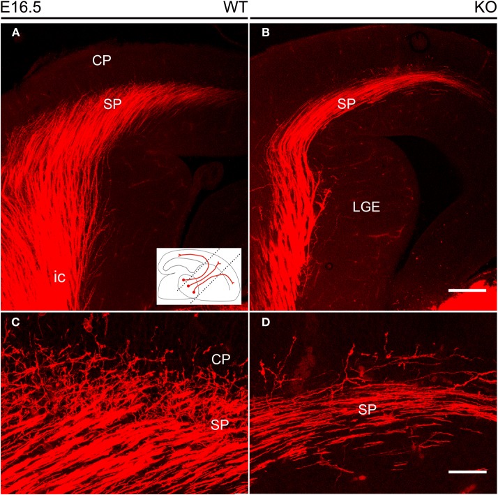
Figure 4.
Thalamocortical axons labeled by DiI inserted into the dorsal thalamus at E16.5. (A,B) DiI-labeled TC axons followed the internal capsule in the ventral forebrain to reach the subplate. The inset shows the orientation of the vertical slices at 45° from the midsagittal plane used to image DiI labeling. In these sections, TC axons in the lateral part of the internal capsule are more anterior than those located medially. (C,D) TC axons started their entry into the cortical plate, but these axonal arborizations were visibly more profuse in wild type (C) than in NCAM null mice (D). Images are maximum projections of confocal optical sections, covering total thicknesses of 29 μm (A,B) or 11 μm (C,D). CP, cortical plate; ic, internal capsule; LGE, lateral ganglionic eminence; SP, subplate. Bars: 200 μm (A,B); 50 μm (C,D).

