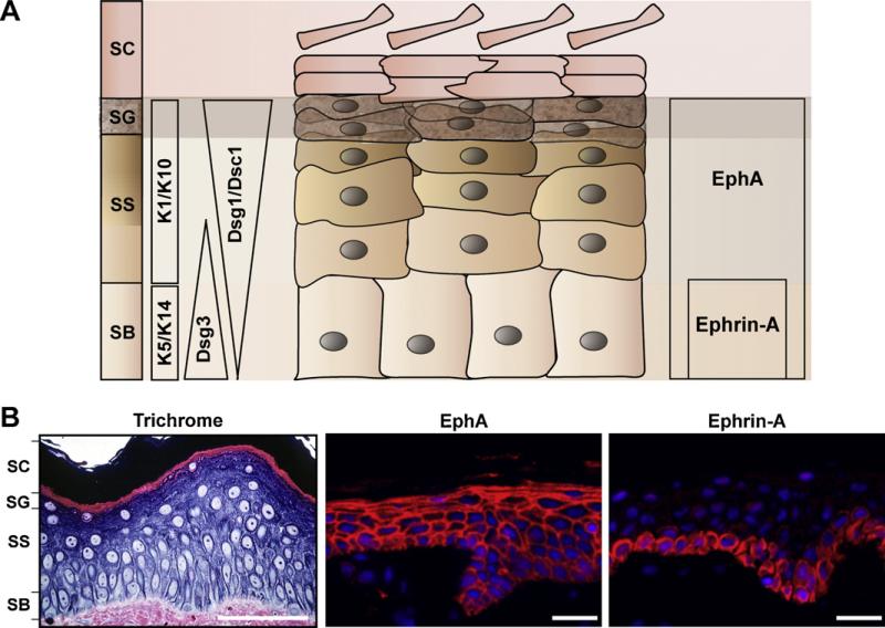
Fig. 2.
Eph receptors and ephrin ligands are compartmentalized in the epidermis. (A) The epidermis consists of four histologically distinct layers which are termed stratum: basale (SB), spinosum (SS), granulosum (SG), and corneum (SC). The epidermal layers are characterized by the expression of several markers of differentiation. Keratin intermediate filaments 5/14 (K5/14) and desmoglein 3 (Dsg3) are present in the basal layer while K1/10, desmoglein 1 (Dsg1), and desmocollin 1 (Dsc1) are concentrated in the more differentiated suprabasal layers of the epidermis. A summary of the distribution of EphA receptors and ephrin-A ligands is presented to the right of the schematic. (B) A 1 μm plastic section of human skin was stained with a trichrome solution (left panel). Black hyphens delineate all four layers of the human epidermis. Human skin samples were immunostained for A class receptors (EphA4) and ligands (ephrin-A1) (right two panels). (Scale bar: 50 μM) Ephrin-A ligands are restricted to the basal layer whereas EphA receptors are expressed throughout all layers of the epidermis.

