
Figure 1.
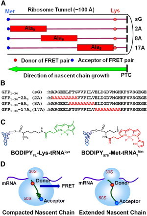
Experimental design of the TR-FRET experiments on poly-A-containing RNCs. (A) Schematic of RNCs in the presence and absence of poly-A segments within the ribosome tunnel. The poly-A fragments are shown in rectangles, with the fluorescent donor and acceptor indicated by red and blue circles, respectively. (B) Sequences of sG, 2A, 8A, and 17A. The parent sequences from GFP are in black and the poly-A inserts are in red. (C) Chemical structures of the fluorescent donor (BODIPYFL) and acceptor (BODIPY576) attached tRNAs. The fluorophores attached to Lys and Met are colored in green (donor) and red (acceptor), respectively. (D) Schematic of 2A with the poly-A insert in a totally helical (left) or extended (right) conformation. The correlation between EFRET and the donor-acceptor separation distance (R) is described by Eq. 9.