
Figure 6.
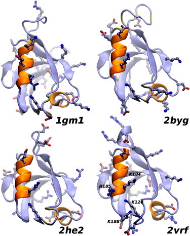
Ribbon representation of four PDZ-domain structures. PDZ2 of PTP-BL (1gm1), second and third PDZ domains of disc large homolog 2, PSD-93 (2byg and 2he2, respectively) and β2S-PDZ (2vrf). In the foreplane are α-helix B and the protein-binding pocket. The side chains of charged residues are shown in all four structures. The amino acids in the characteristic cluster of arginine and lysine residues in β2S-PDZ are labeled.