
Figure 1.
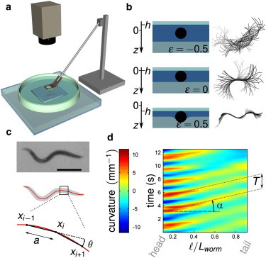
Experimental framework. (a) Setup. A worm is confined between an agar gel and a glass plate that can be moved vertically. Movies are recorded with a charge-coupled device camera through a stereo microscope (see Materials and Methods). (b) (Left) Cross sections of the confined worm, for different values of ϵ; (right) corresponding superpositions of the worm midline over 2 s. (c) Image analysis. (Top) Raw image (scale bar 0.5 mm); (center) thresholded image (gray) and skeletonization (red line); (bottom) the body curvature is computed as κ = θ/a. (d) Spatiotemporal plots of the curvature versus a nondimensional curvilinear coordinate from head to tail (ℓ/Lworm).