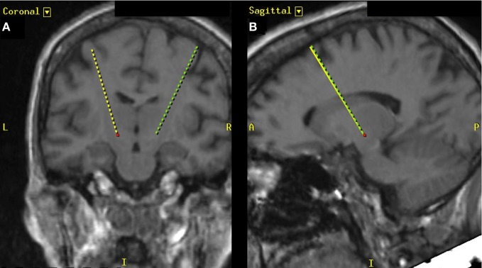
Figure 1.
T2-weighted MR image depicting the targeting trajectory (dashed lines) for the placement of deep brain electrodes. (A) Depiction of electrode trajectories in the coronal plane and (B) depicts the electrode trajectory in the sagittal plane. The red dot indicates the position of the left STN.

