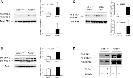
Fig. 4.
Western blot analysis of ERK and JNK phosphorylation in the lungs of mice. A–B: Western blot analysis of phospho-ERK (ph-ERK) and phospho-JNK (ph-JNK) in the lungs of Apoe−/− mice after 10 wk of WTD. Densitometric analysis of the signal was performed to determine ph-ERK/total ERK and JNK/actin ratios. C: Western blot analysis of ph-ERK in the lungs of Ldlr−/− mice fed a WTD for 10 wk. D: peritoneal macrophages from Apoe−/− and Apoe+/+ treated with TLR4 ligand (100 ng/ml) and oxidized LDL (100 μg/ml). Activation of ERK in Apoe−/− macrophages treated with oxidized LDL was augmented by the addition of TLR4 ligand.

