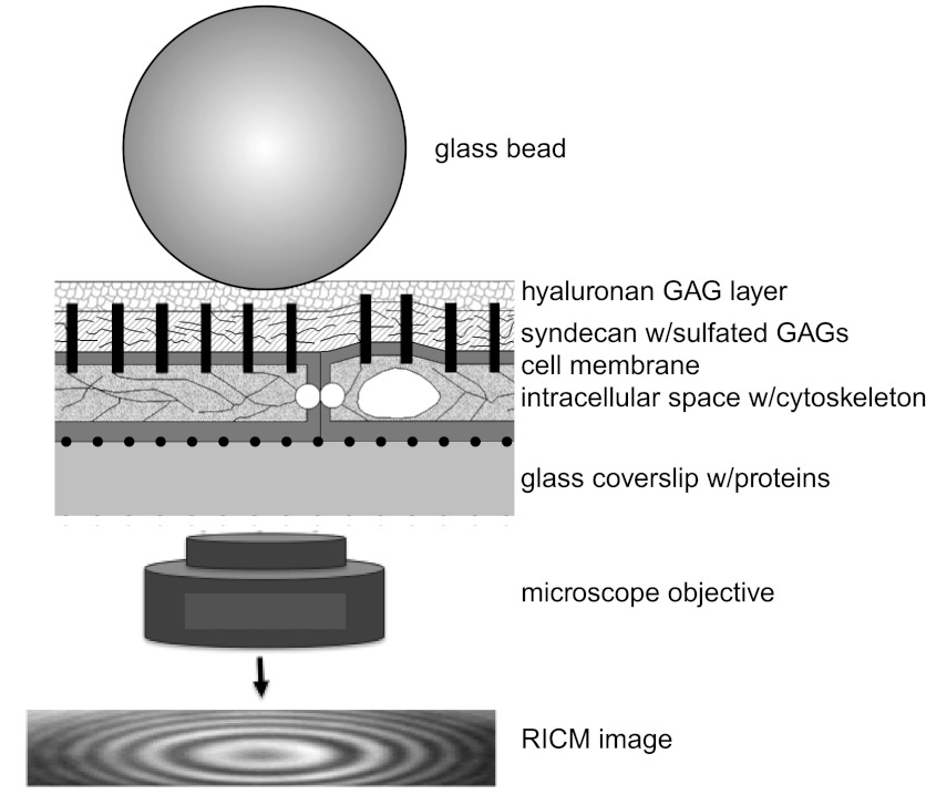
Fig. 1.
Schematic of reflectance interference contrast microscopy (RICM) bead probe placed on the top of bovine lung microvascular endothelial cell (BLMVEC) glycocalyx (not drawn to scale). Monochromatic light reflected from bead and coverslip surface constructively interferes to create an interference pattern in the RICM image that is captured by microscope objective and CCD camera. The interference pattern changes when the distance between the bead and the coverslip change. GAG, glycosaminoglycan.

