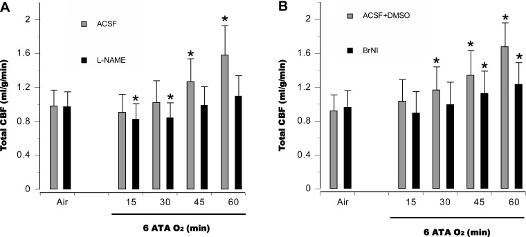
Fig. 3.
Cerebral hemodynamics. In anesthetized control animals, total cerebral blood flow (tCBF) was relatively stable for the first 30 min at 6 ATA and then increased, peaking as EEG spikes appeared. In anesthetized animals pretreated with l-NAME (A), tCBF was less than in control animals for the first 30 min, and minor increases were observed over the rest of hyperoxic exposure. In the BrNI group (B), tCBF within 30 min of HBO2 did not differ from baseline; however, subsequent increases were significant, but less pronounced than in control animals. *P < 0.05 vs. air in A and B.

