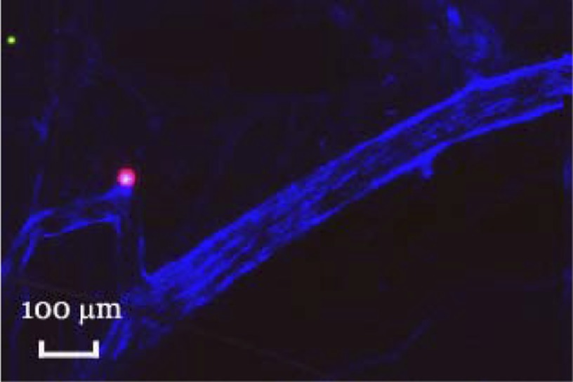
Fig. 3.
Transverse section through the base of a rat brain. One million 15 (green)- and 33 (red)-μm spheres were injected into the superior vena cava, followed by 100 μl Evan's Blue dye to label the remaining patent vasculature. The brain was removed, embedded in Tissue Tek, sliced, and imaged confocally. Note that embolization by the 33-μm microsphere has disrupted perfusion.

