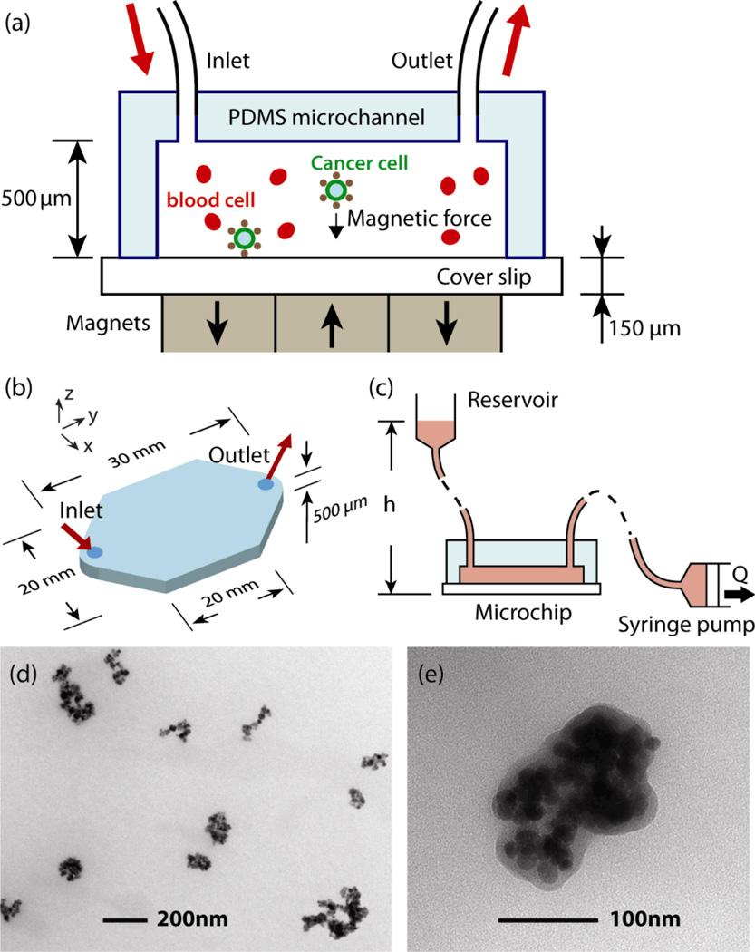
Fig. 1.
Microchip design for immunomagnetic detection of cancer cell. (a) Schematic showing the principle of operation. CTCs in blood are labelled with EpCAM functionalized Fe3O4 magnetic nanoparticles, and captured by the magnetic field as the blood flows through the microchannel. (b) Dimensions of the microchannel. (c) Schematic of the pneumatic flow system. The flow rate is regulated by the syringe pump from 2.5–10 mL/hour, which draws the blood rather than pushing it to minimize the inside pressure of the chamber. (d–e) TEM images of Fe3O4 magnetic nanoparticles.

