
Figure 3.
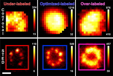
Effect of labeling density in STED images of Cep164 in MTECs. (Top panel) Confocal and (bottom panel) STED images of Cep164 in MTECs immunostained with increasing concentrations of primary antibody and fixed concentration of secondary antibody. With increasing primary antibody concentration, the number of observed clusters increases until saturation at nine clusters. Overlabeling with antibody blurs the Cep164 centriole substructure. Pixel intensities are represented in the color bars. Scale bar: 200 nm.