
Figure 5.
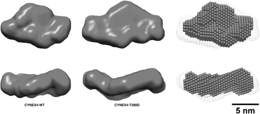
Modeling of CYNEX4 biosensor constructs. Ab initio bead models of CYNEX4 (left) and the phosphorylation mimic T266D (middle), from the program DAMMIF, are shown as solid surfaces. On the right is a best-superposition from SUPCOMB13 of the CYNEX4-wt (spheres) and the T266D (mesh surface) bead models. The lower structures in each panel are rotated through 90° along the horizontal axis.