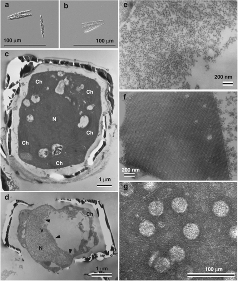
Figure 2.
Thalassionema nitzschioides AR-TN01 isolated from surface water in Ariake Sound, Japan. (a) Optical micrograph of intact cells. (b) Cultures inoculated with TnitDNAV at 15 d.p.i.. Transmission electron micrographs of ultra-thin sections of T. nitzschioides and negatively stained TnitDNAV particles. (c) Healthy cell. (d, e and f) Cells infected with TnitDNAV at 8 d.p.i. (d) Degraded host cytoplasm and chloroplast. (e) Higher magnification of randomly aggregated VLPs in the host nucleus in panel d. (f) Para-crystalline array aggregation of VLPs in an infected host nucleus. (g) Negatively stained TnitDNAV particles in purified culture lysate. Arrows denote the accumulation of VLPs. N, nucleus; Ch, chloroplast; V, virus-like particles.

