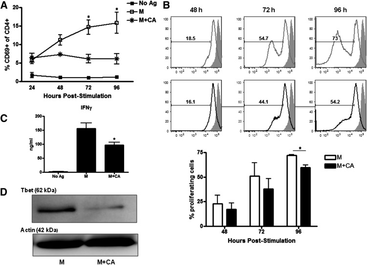
FIG. 2.
Redox modulation promotes a reduction in T-cell activation and effector function. A: BDC-2.5.TCR.Tg splenocytes were left untreated or stimulated with M ± CA. At 24–96 h, cells were stained for and gated on CD4+ cells, and CD69 was analyzed by flow cytometry (n = 3 independent experiments). B: Carboxyfluorescein succinimidyl ester (CFSE)-labeled splenocytes were treated with M ± CA. At 48–96 h, cells were stained and gated on CD4+CFSE+ cells. Gray lines, M; black lines, M + CA; filled histograms, CFSE+ cells at time point 0; % proliferating cells, average of three independent experiments. C: Supernatants from 96-h cultures were used in an IFN-γ ELISA (n = 3 independent experiments performed in triplicate). D: Whole-cell lysates from 96-h cultures were probed for Tbet by Western blot. Actin was probed as a loading control. Data are representative of three independent experiments. *P < 0.05.

