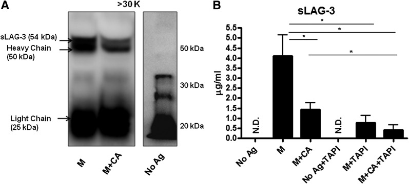
FIG. 5.
sLAG-3 protein is decreased after CA treatment. A: Supernatants from BDC-2.5.TCR.Tg splenocytes left untreated or stimulated with M ± CA for 48 h were concentrated using Amicon Ultra Centrifugal Filters at a 30-kDa (K) cutoff. The >30-kDa portion was then immunoprecipitated with anti–LAG-3 antibody, separated on an SDS-PAGE gel, and probed for LAG-3 by Western blot. Data representative of three independent experiments. B: BDC-2.5.TCR.Tg splenocytes were left untreated or stimulated with M ± CA or TAPI-1 for 72 h. Supernatants were collected and used in sLAG-3 ELISAs. Graph shows the average of four independent experiments performed in triplicate. *P < 0.05. Ag, antigen; N.D., none detected.

