FIG. 6.
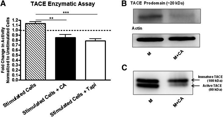
Redox modulation diminishes active TACE levels and enzymatic function. A: BDC-2.5.TCR.Tg splenocytes were stimulated with M ± CA ± TAPI for 24 h and supplemented with TACE-specific fluorogenic substrate. Fluorescence was measured at 6 h post substrate addition. The fold change in activity was calculated by stimulated/unstimulated vs. stimulated + CA/unstimulated vs. stimulated + TAPI/unstimulated cells. Graph shows the average of three independent experiments performed in triplicate. **P < 0.005, ***P < 0.0005. B and C: BDC-2.5.TCR.Tg splenocytes were stimulated with M ± CA for 72 h and probed for TACE by Western blot. Whole-cell lysates were used in B. Actin was probed as a loading control. Membrane lysates were used in C. Data are representative of three independent experiments.

