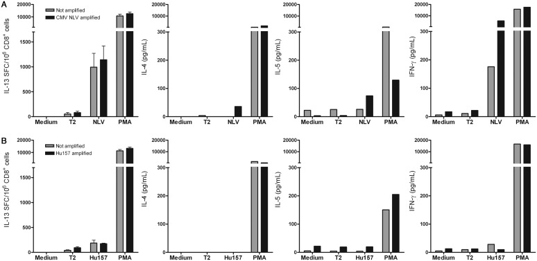
Fig. 2.
Secretion of type 2 cytokines and IFN-γ by CD8+ T cells after stimulation with cDCs. CD8+ T cells were tested for secretion of type 2 cytokines and IFN-γ in response to peptide-pulsed T2 cells after stimulation with peptide-pulsed cDCs (amplified, black bars) or cDCs without peptides (not amplified, grey bars). Upper panels (A) show the results of T cells obtained from a CMV-seropositive healthy donor, lower panels (B) show the results for Hu-PNS patient no. 7. CD8+ T cells were tested in medium and against T2 cells (T2), T2 cells pulsed with the CMV pp65-derived peptide NLV, T2 cells pulsed with the HuD-derived peptide Hu157, or T2 cells that were added simultaneously with PMA plus ionomycin (PMA). Panels on the left show the numbers of IL-13 SFC/106 CD8+ T cells, the other panels show cytokine concentrations in culture supernatants (pg/mL) of IL-4, IL-5, and IFN-γ. The CD8+ T cells of the CMV-seropositive healthy donor (upper panels) secreted both IL-13 and IFN-γ. CD8+ T cells of Hu-PNS patient no. 7 (lower panels) did not secrete the type 2 cytokines IL-4, IL-5, or IL-13 or IFN- γ. Abbreviations: cDCs, conventionally generated dendritic cells; CMV, cytomegalovirus; PMA, phorbol myristate acetate plus ionomycin; IL, interleukin; PNS, paraneoplastic neurological syndromes.

