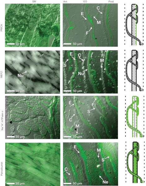
Fig. 4.
Protein localization in EO and SM. Immunohistochemistry was performed using primary antibodies for four additional proteins: plasma membrane Ca2+-ATPase (PMCA), myocyte enhancing factor 2 (MEF2), the α subunit of the Na+/K+-ATPase and parvalbumin, which are listed in Table 3. The images presented in this figure are overlays of DIC and fluorescence images (see Materials and methods). Illustrations are provided to summarize the localization of each protein. For all images, anterior is left, posterior is right. Abbreviations: E, electrocytes; C, connective tissue septa; Nu, nuclei; F, myofilament material between the anterior and posterior membranes; M, microstalklets; P, penetrations; S, stalk; Ne, motor neuron. See Fig. 3 for an overview of anatomical features in the EO.

