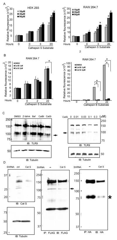
Figure 4.
Generation of sTLR9 is regulated by cathepsin S. (A) The lysates from RAW 264.7 and HEK 293 cells were incubated with 10, 30, or 90 μM of quenched fluorescent cathepsin S substrate. Some lysates were incubated without substrate to determine background fluorescence (0, no substrate). At 0, 3, and 20 hours fluorescence was measured. Increased fluorescence indicated cleavage of the substrate. (B) Cathepsin S and cathepsin B activity were measured as in (A) except the RAW 264.7 cells were incubated with DMSO control, 2μM cathepsin S inhibitor or 1μM cathepsin B inhibitor and fluorescence was measured at 0, 2, and 22 hours. (C) HEK293 cells stably expressing TLR9-YFP were incubated for six hours with 10μM Z-FA-fmk, 100nM Bafilomycin A1, 10μM cathepsin B inhibitor or 30nM cathepsin S inhibitor (left panel), or with different concentrations of cathepsin S inhibitor (right panel). Whole cell lysates were resolved by SDS-PAGE, transferred to nitrocellulose, and immunoblotted for TLR9 (N-Term) and tubulin. ➛ sTLR9. (D) Whole cell lysates of HEK 293 cells stably tranduced with control or cathepsin S shRNA were resolved by SDS-PAGE and analyzed by immunoblot analysis with anti-cathepsin S, or tubulin (left panel). Cathepsin S shRNA or control HEK 293 cells were transfected with UNC93B1 and flag-mTLR9 (middle panel) or UNC93B1 and mTLR9-HA (right panel). The lysates were immunoprecipitated for either FLAG (middle panel) or HA (right panel). The immunoprecipitates were resolved by SDS-PAGE gel, transferred to nitrocellulose, and immunoblotted for either FLAG (N-Term, middle panel) or HA (C-Term, right panel). Data are representative of three independent experiments.

