
Figure 5.
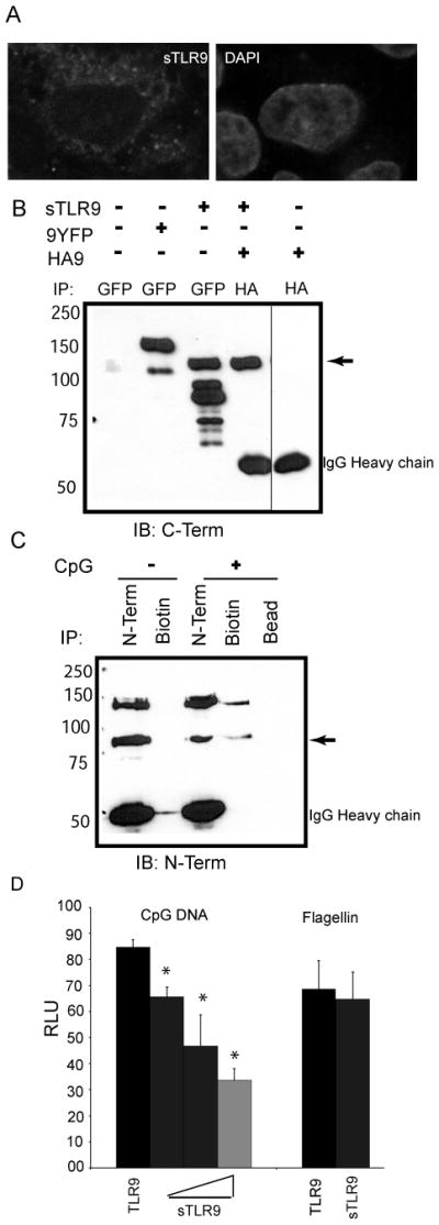
sTLR9 binds to CpG DNA and negatively regulates TLR9 signaling. (A) HeLa cells were transfected with sTLR9 tagged with YFP at the C-terminal end, fixed, mounted in anti-fade with DAPI, and imaged by confocal laser scanning microscopy. (B) HEK 293 cells were transfected with full length TLR9-YFP (9YFP), sTLR9-YFP (sTLR9), N-terminally HA tagged-TLR9 (HA9) or sTLR9-YFP and HA-TLR9. Lysates were immunoprecipitated for GFP/YFP or HA as indicated and assayed by immunoblotting for GFP/YFP. ➛ sTLR9-YFP. (C) A human B cell line was treated with media (−) or 3′ biotinylated CpG DNA (+) for 15 min, lysed, and immunoprecipitated for TLR9 (N-Term), biotin, or incubated with beads alone (Bead). Immunoprecipitates were analyzed by immunoblotting for TLR9 (N-term). ➛ sTLR9-YFP. (D) HEK 293 cells were transfected with a plasmid encoding full length TLR9 either without (black bar) or with increasing concentrations of a plasmid encoding TLR9 amino acids 1–723 (sTLR9) (grey bars), plus an NF-kB-luciferase reporter. After overnight stimulation with 2 μg/mL CpG DNA, or 100 ng/mL flagellin as a control, cells were lysed, and assayed for luciferase activity. Data are representative of three independent experiments.