
Figure 2. DE-DM cell in vitro co-culture.
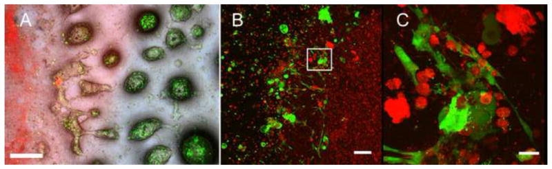
A) Labeled DE and DM cells still exhibited strong fluorescence after 7 days in vitro culture. Merged bright field fluorescent images: green DE cells in Matrigel; red DM cells in Collagen. B) Confocal imaging revealed polarized DE and DM cell interactions along the cell seeded gels interface. C) High magnification view of B (white box). Scale bar=500 μm (A), 100 μm (B), and 20 μm (C).