
Figure 3. DE-DM construct.
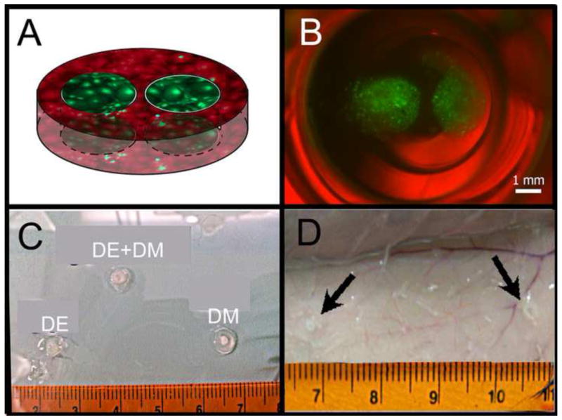
A) Schematic of DE-DM construct containing green labeled DE cells and red labeled DM cells. B) Light micrograph image of fluorescent DE-DM construct after 2 days in vitro culture. Merged bright field photo and fluorescent images: green DE cells in Matrigel; red DM cells in Collagen. C) Dental cell seeded constructs prior to implantation. DE-DM and DM only implants maintained their shape, while the DE collagen implant was very soft. D) Subcutaneous implant after 4 weeks in vivo (arrows).