
Figure 4. Staged Hematoxylin and eosin (H&E) stained in vitro cultured DE-DM constructs.
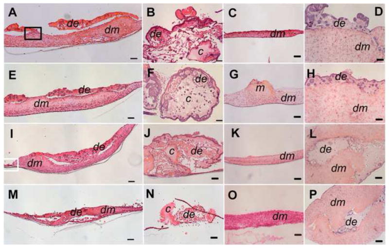
Porcine DE and human DM cell construct (A, E, I, M). Porcine DE cell construct (B, F, J, N). Human DM cell construct (C, G, K, O). Porcine DE and porcine DM cell constructs (D, H, L, P). 1 wk (A–D), 2 wk (E–H), 3 wk ((I–L), and 4 wk (M–P) in vitro cultured constructs. In all constructs, DE cells formed rosette and tubular like structures (arrows). Matrigel and collagen gels showed significant degradation by 4 wks in vitro. Abbreviations: de: DE cells with Matrigel; dm: DM cells with Collagen gel; m: Matrigel without DE cells; c: Collagen gel without DM cells. Scale bar=100 μm.