
Figure 6. IHC analyses of 2 wk in vitro cultured DE-DM structures revealed cell differentiation and basal membrane formation.
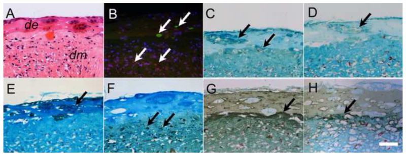
A) H&E stained DE-DM layer interface. B) Serial sectioned construct revealed fluorescent stained DE (green) and DM (red) cells. C) DSPP expression in both DE rosettes and DM layer. D) AM expression was detected only in DE cell layer. E) CK-14 positive DE cell layer. (F) Vimentum was detected only in the DM cell layer (F). Strong Laminin (G) and collagen IV (H) expression was detected at the DE-DM border. In all panels, arrows indicate positive expression. Scale bar=20 μm.