
Figure 8. Human dental pulp cells and mineralized tissues in 4 wk DM in vivo constructs.
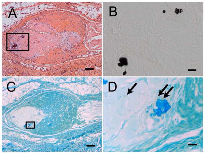
A) H&E stained DE-DM cell construct revealed mineralized tissue formation. B) High magnification view of Von Kossa stained area boxed in Panel A. (C) Human dental pulp cells expressing human mitochondrial protein were located adjacent to calcified tissues (D, arrows). Scale bar = 100 μm (A, C), and 20 μm (B, D).