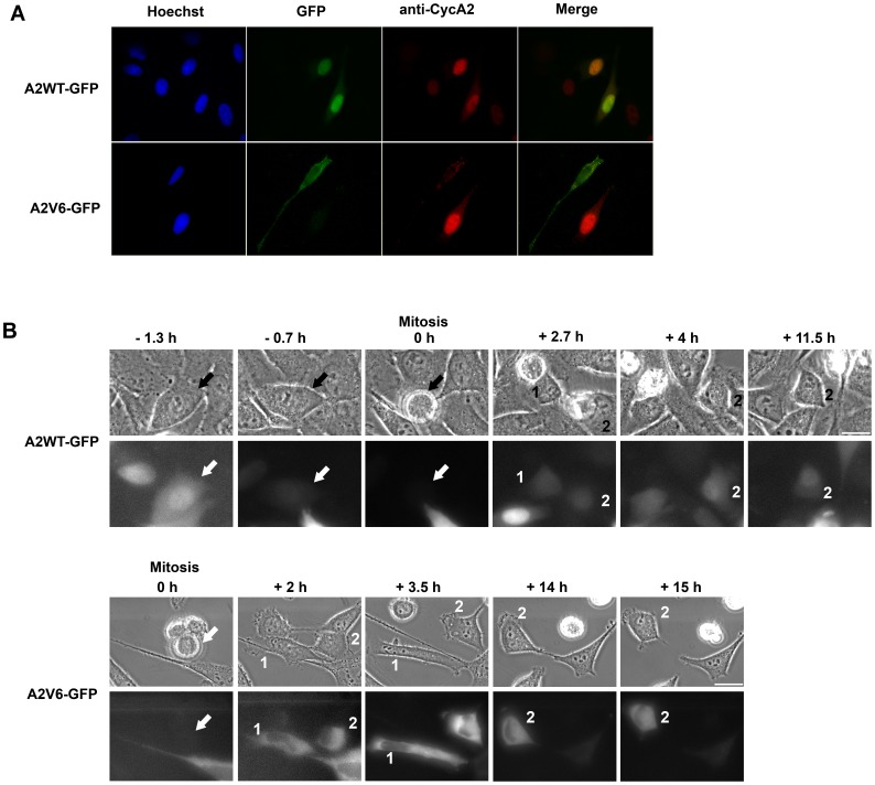
Figure 2. Cytoplasmic localization of A2V6-GFP during the cell cycle.
HeLa cells were transfected with wild-type cyclin A2-GFP (A2WT-GFP) or Intron 6-retaining cyclin A2-GFP (A2V6-GFP) vectors. (A) Immunofluorescence images of transfected HeLa cells using anti-GFP (green) and anti-cyclin A2 (red) antibodies. Nuclei (blue) were counterstained with Hoechst dye 33258. The A2V6-GFP protein is located in the cytoplasm of transfected cells. No endogenous cyclin A2 is detected in cells expressing the A2V6 protein. (B) Time-lapse imaging of A2WT- and A2V6-GFP during a cell cycle. Upper panel: Phase contrast microscopy. Lower panel: Fluorescence microscopy. Arrow: parent cell. Labels 1 and 2: daughter cells. Wild-type cyclin A2-GFP is accumulated in G2 nuclei (−1.3 h), degraded in mitosis and reappears mostly in the nuclei in S phase, like endogenous cyclin A2. In contrast, A2V6-GFP remains in the cytoplasm throughout the cell cycle. Scale bar: 20 µm.

