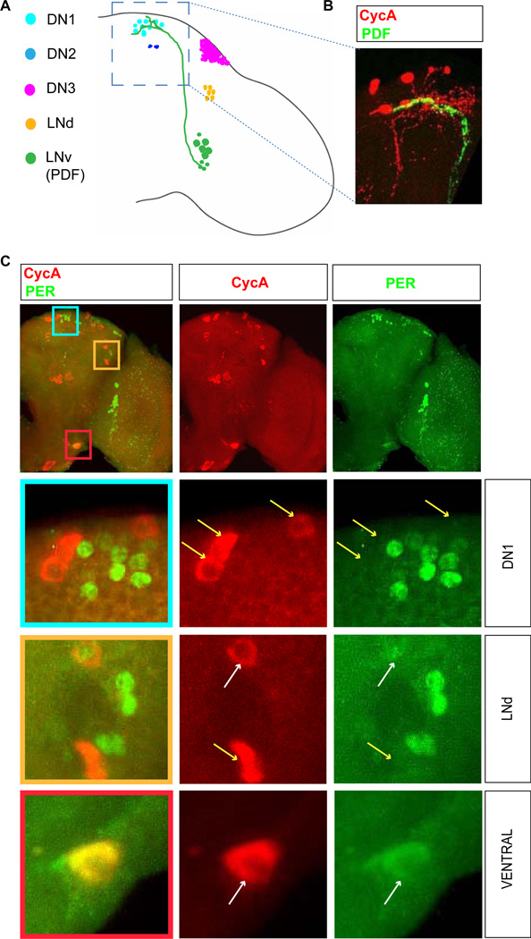
Figure 4. Association of CycA neurons and circadian clock neurons in the brain.
(A) A schematic representation of an adult brain with known groups of clock neurons indicated by different colors. LNv neurons project to the dorsal area of the brain where DN1 clock neurons are found. (B) The bodies and arborizations of the dorsal CycA neurons (red) are found in the vicinity of the LNv neuronal termini (green, marked by the expression of neuropeptide PDF). (C) Immunostaining for CycA (red) and a core circadian clock component Period which marks circadian clock neurons (PER, green). The bottom three panels show higher magnification images indicated by different color squares in the upper panel. The labels “DN1” and “LNd” refer to the types of circadian neurons found in these respective areas, while “ventral” denotes an area that has no known clock neurons. In different regions of the brain, cells labeled for CycA and PER cluster (DN1 region), cluster and partially overlap (LNd region), or overlap (ventral region). White and yellow arrows point to overlapping and non-overlapping cells, respectively. For simplicity, only one brain hemisphere is shown. The brains shown were fixed at Zeitgeber time 2 (ZT2, the time point at which levels of PER are high).

