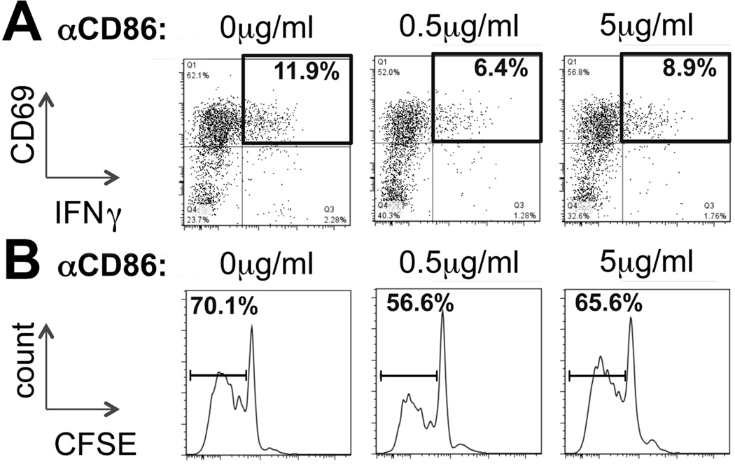
Figure 4. Hepatic costimulation of T-lymphocytes in BA is mediated by CD86.
(A) Hepatic pan-DCs were purified from RRV infected mice at 7dpi and cultured with CFSE-labeled naïve CD8 cells from non-infected neonatal mice in presence of various concentrations of antibodies against CD86. CD8 activation, correlating with expression of CD69 and IFNγ, was determined by flow cytometry after 72 hours of culture. The numbers in representative dot plots denote frequencies of CD69+IFNγ+ cells gated on CD8 cells. (B) Proliferation of CD8 cells was determined in a CFSE dilution assay. The numbers above the intervals in the histograms denote percentages of CD8 cells with decreased cellular CFSE content, correlating with the frequency of proliferating CD8 cells in the co-culture. Plots and histograms are representative for 2 independent experiments.

