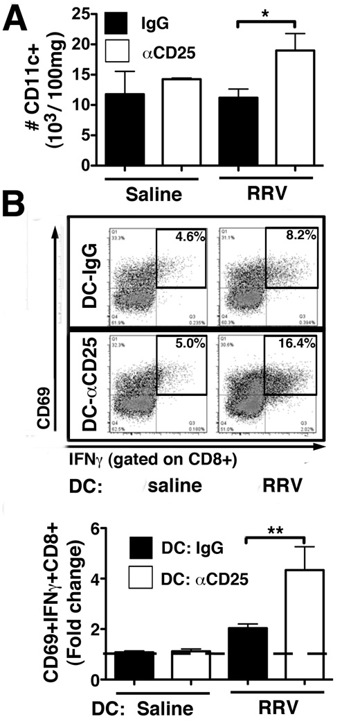
Figure 8. Treg-depletion enhances stimulatory capacity of hepatic DCs.
(A) Hepatic MNCs were purified from αCD25- and IgG-treated mice at 12 dpi after RRV (or saline) injection on day 8 of life. Total numbers of hepatic CD11c+ DCs were determined by flow cytometry. Values are displayed as mean+SEM with *p<0.05 in unpaired t test (n=2–4 mice per group). (B) Purified hepatic pan-DCs from the 4 experimental groups were co-cultured with CD8 cells from non-infected neonatal mice for 48 hours. CD8-activation was determined by flow cytometry and numbers in representative dot plots denote frequencies of CD69+IFNγ+ gated CD8 cells. The fold change of %CD69+IFNγ+CD8+ in DC/CD8 co-cultures compared to CD8 cells cultured without DCs (denoted by the dashed line) is displayed in the bar graph. Values are expressed as mean+SEM of fold changes with 6–9 pooled livers per DC condition in 3 independent experiments with ** p<0.01 in unpaired t test.

