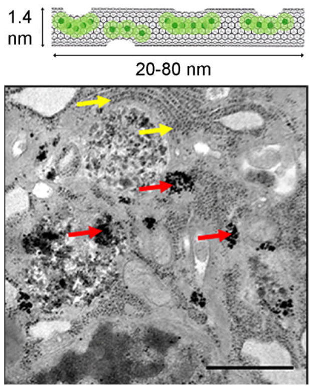
FIGURE 4.
(A). A representative illustration of gadonanotubes. Clusters of internally-loaded Gd3+ ions are located at defect sites along the nanocapsule sidewalls. (B). TEM images of a gadonanotube-labeled MSC. Red arrows point to gadonanotube aggregates in the cytoplasm. Yellow arrows point to ribosomes of the endoplasmic reticulum. Scale bar = 1 μm. (Reproduced with permission from Ref (47). Copyright 2011 Elsevier B.V.).

