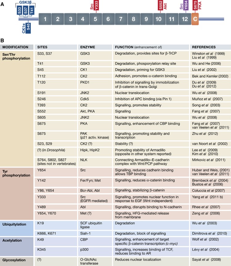
Figure 4.
The functional output of β-catenin is affected by post-translational modifications. (A) A representative scheme showing the sites where β-catenin is phosphorylated: those in blue promote its degradation, those in red and purple enhance the signalling activity. The Y654 site (purple) was experimentally validated in a mouse model in vivo. The protein kinases are denoted that promote each phosphorylation. (B) Table summarizing possible post-translational modifications of β-catenin with their functional consequences.

