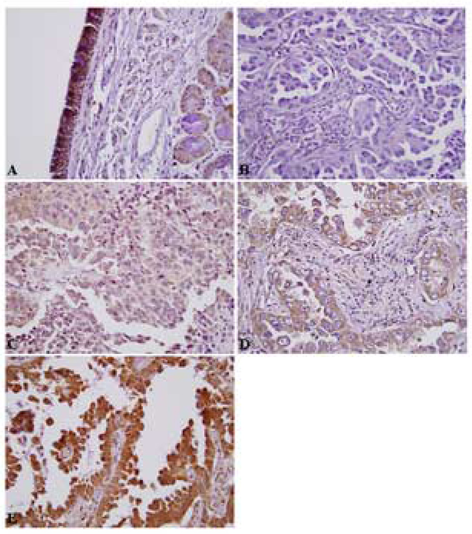
Figure 1.
Immunohistochemical patterns for C/EBPα expression in E3590. Representative images: A) Normal bronchial mucosa with strong C/EBPa staining of bronchial epithelium (3+). B) Lack of C/EBPα staining (0) within tumor cells, C) Faint C/EBPα staining (1+), D) Moderate staining (2+), E) Strong C/EBPα staining within tumor cells (3+, which is comparable to that of the normal bronchial epithelium). (A, 100X; B, C, D and E, 400X).

