Figure 6.
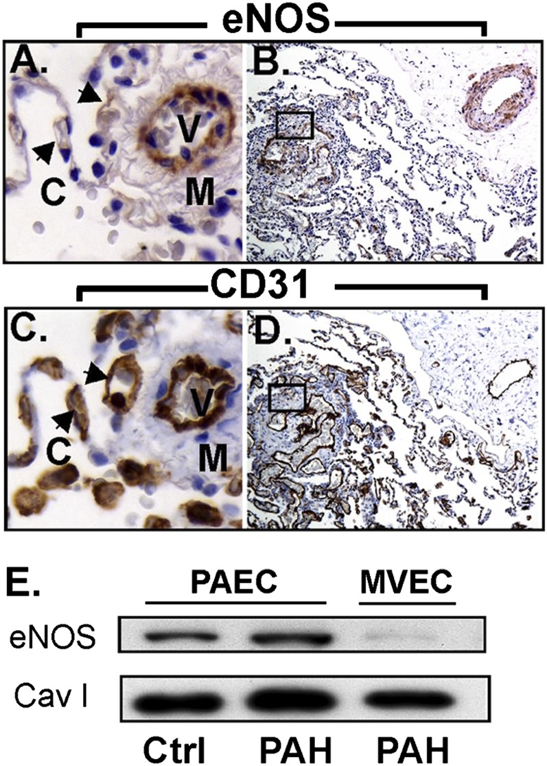
Endothelial nitric oxide synthase (eNOS) expression in PAECs and MVECs. (A, B) Immunohistochemistry shows strong eNOS staining in the muscular vessels where the staining in alveolar capillaries (arrows) is absent. The plexiform lesion (A) is lined with endothelial cells that are mostly positive for eNOS, but some cells are negative. (C, D) Sequential section confirms endothelial cells by CD31 and in plexiform lesions. (E) Western blot analysis shows that control and PAH PAECs express eNOS, whereas MVECs have undetectable or weak expression of eNOS. C = capillaries; M = smooth muscle; V = vessel.

