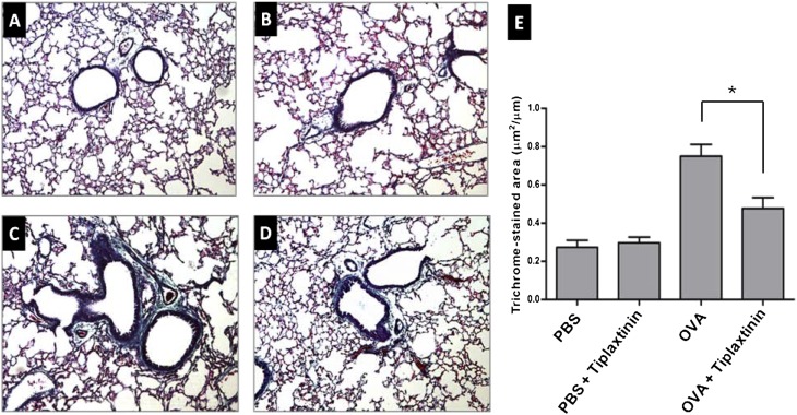
Figure 5.
Effect of tiplaxtinin treatment on collagen deposition. For assessment of collagen deposition, tissue sections of lungs from mice were stained with Gomori trichrome (left panels) and quantified in trichrome-stained area/length of bronchial basal membrane by microscopy and Image J (E, right panel). Mice were nebulized with PBS (A and B) or OVA (C and D), and treated with tiplaxtinin during the nebulization (B and D). The data in the histogram (E) are means ± SEM (n = 8). *P < 0.05 between groups.

