Figure 4.
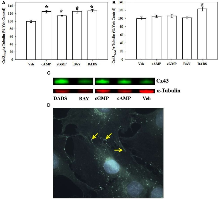
Total Cx43 expression in VSMCs. (A) In-cell Western blotting for total Cx43 expression in rat primary VSMC homogenates. Incubation of rat primary VSMCs for 270 min in the presence of 8Br-cAMP (100 μM), 8Br-cGMP (100 μM), BAY (100 nM), or DADS (50 μM) significantly stimulated total Cx43 expression normalized to α-tubulin. Data are from two independent experiments each performed in triplicate. (B) After 24-h incubation the stimulation of Cx43 was significantly increased only after DADS treatment with no observable changes for the 8Br-cAMP, 8Br-cGMP, or BAY treatment groups compared with vehicle controls. For these experiments n = 6 for each treatment. Student–Newman–Keuls method for multiple comparisons following one-way ANOVA was used. *p < 0.05 compared with normalized vehicle controls. (C) A representative Western blot showing total Cx43 in green and α-tubulin in red using IR-labeled antibodies. These data were obtained from a single Western blot with a column (unrelated to the current study) removed for clarity. (D) Photomicrograph of confluent VSMCs following immunocytochemical staining for total Cx43. Punctate appearance of Cx43 indicating the presence of gap junctions is predominantly at cell-to-cell contacts (indicated by arrows).

