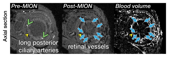
Figure 2a:
Three-dimensional MR microangiography and volume rendering reveal vascular morphologic features of a rat eye. (a) Oblique axial MR images (26/4.3) perpendicular to the optic nerve head show the retinal vessels (blue arrows) branching from the central retinal artery, with LPCAs (green arrowheads). (b) Oblique sagittal MR views (26/4.3) parallel to the optic nerve head of the eye depict the central retinal artery, choroid (yellow arrowheads), and the retinal vessels (blue arrows). (c) Volume-rendered 3D images show eyeball at left and vascular structure at right without MION injection. x = Readout direction, y = first phase-encoding direction, z = second phase-encoding direction. (d) Volume-rendered 3D image of the ocular BV. Post-MION = after MION injection, Pre-MION = before MION injection, green arrowheads = LPCAs, red arrows = posterior ciliary artery.

