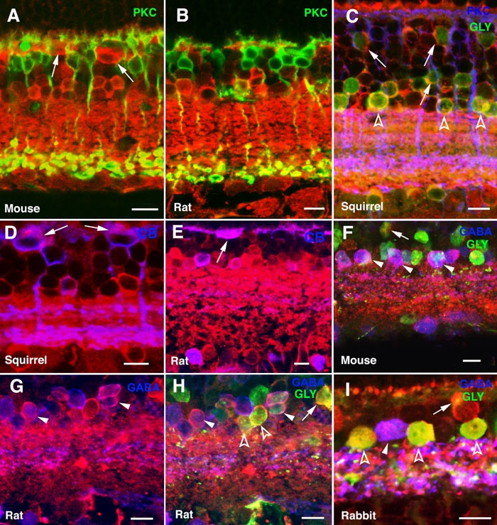
Figure 5.

Expression of α-synuclein in horizontal, bipolar, and amacrine cells. Shown are immunolabeled sections of (A, F) mouse, (B, E, G, H) rat, (C, D) squirrel, and (I) rabbit retinas. α-Synuclein immunostained red in all panels. Single, double, or triple labelings were carried out for the indicated retinal neuronal markers. Protein kinase C (PKC) stained green in panels A and B and blue in C. Glycine (GLY) stained green in panels C, F, H and I. Calbindin (CB) stained blue in panels D and E, as did GABA in panels F-I. Arrows point to horizontal cells in A, D and E, and to glycinergic bipolar cells in panels C, F, H, and I. Open arrowheads point to glycinergic amacrines (panels C, H, I) and closed arrowheads to GABAergic amacrines (panels F-I). Each bars equals 10 μm.
