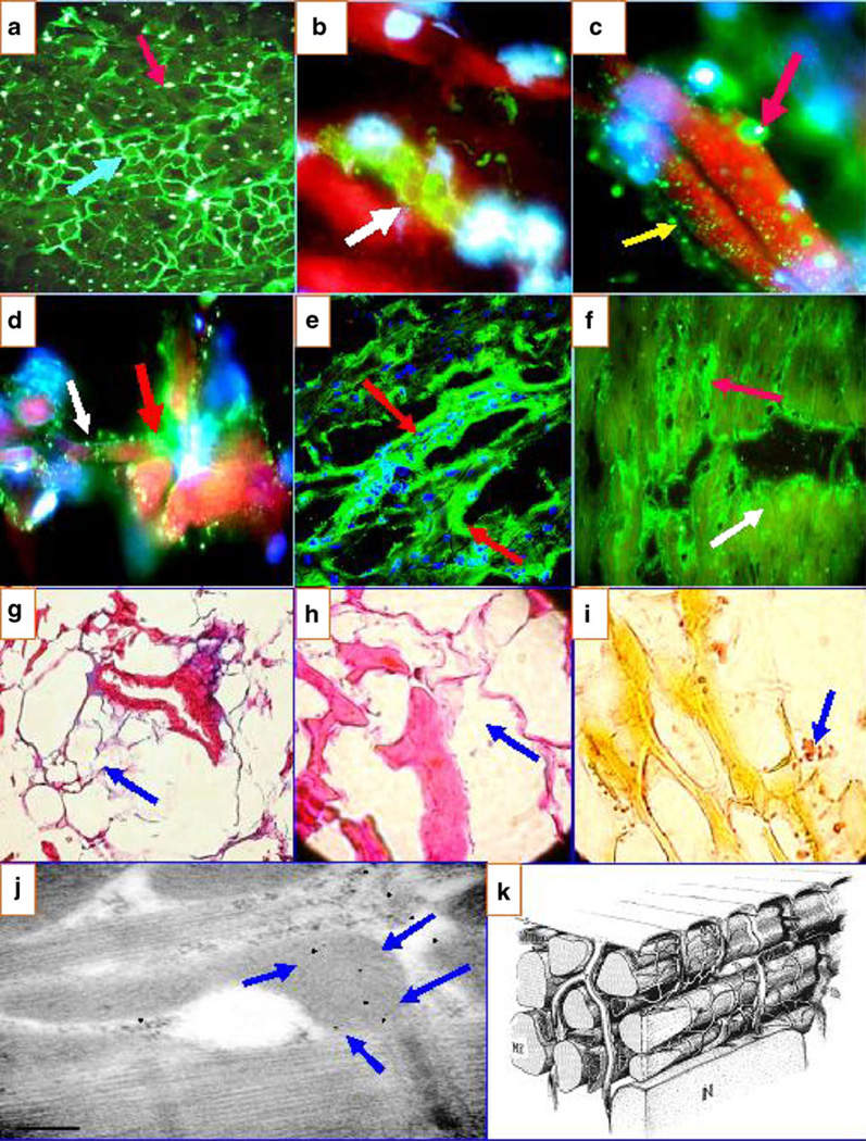
Fig. 1.
a Anti-human fibrinogen antibody conjugated with FITC highlight the intercellular reactivity to the area composita of the heart (blue light arrow). b Positivity against rounded structures in proximity to the TTS using FITC-conjugated anti-human IgG antibody (green reactivity) (red arrow). c Two types of reactivity using FITC-conjugated anti-human IgG antibodies: one is observed against the larger round structures (reactivity in green; red arrow) and another directed against smaller structures (yellow arrow). d The same antibody as in c, showing reactivity to the TTS (green staining) (red arrow) and to the smaller structures (white arrow). e a CFM image demonstrating the reactivity to the TTS (green staining; red arrows) using FITC-conjugated anti-human IgG antibody. f Similar to e but at lower magnification. The red arrow shows the reactivity to the TTS, and the white arrow shows the reactivity to the smaller, round structures (faint yellow staining). In addition, in g (Masson’s trichrome), h (H&E), and i (Verhoeff elastin), we show some representative pictures of the loss of collagen and elastin fibers and some replacement with lipomatous tissue, respectively (blue arrows). j IEM highlighting positive staining against the heart mitochondria in a patient with El Bagre-EPF (blue arrows). The other reactivity (small black dots) is against cell junctions. k A model of myocardial fibers, the components of the sarcoplasmic reticulum and their relationship with the TTS

