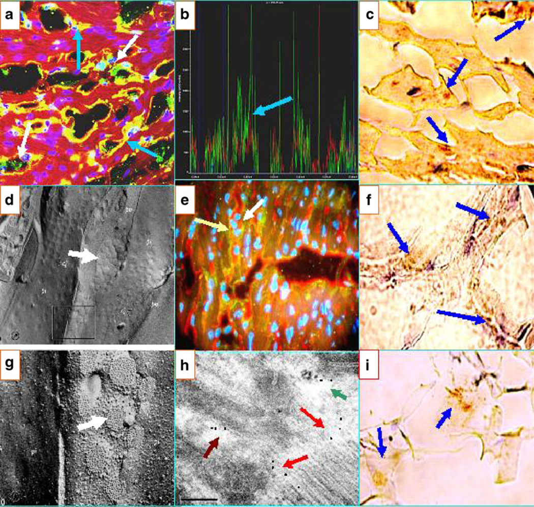
Fig. 2.
a Confocal image showing positivity against the area composita of the heart. The white arrows show green dots representing FITC-conjugated anti-human IgG antibody autoreactivity. The blue arrows show autoreactivity (yellow staining) to the TTS. The nuclei are counterstained in purple with DAPI. b CFM colocalization of the a peaks of fluorescence, indicating that the antibodies to DP I–II (red staining) colocalize with the patient antibodies (green staining; blue arrow). The faint blue peaks represent the DAPI nuclear counterstaining. c IHC showing positive staining of one El Bagre-EPF patient necropsy heart with anti-human IgE antibody (blue arrows; dark staining). e DIF showing colocalization of antibodies to Connexin 43 in red (white arrow) with an El Bagre-EPF patient serum (green staining; light yellow arrow). d, g Pictures from tissue fracture electron microscopy of the area composita (white arrows). f IHC showing positive staining within the heart using anti-human fibrinogen antibody (blue arrows). h An IEM photograph of the heart showing El Bagre-EPF serum labeled with 10 nm gold-conjugated protein A antibodies, reacting with multiple heart structures including the area composita (maroon arrow; black dots), an interface area of the desmosomes and adherens junctions (red arrows), and an area that resembles either gap and/or tight junctions (green arrow) (100KX). i An IHC stain of El Bagre-EPF patient heart, staining positive with complement/C1q

