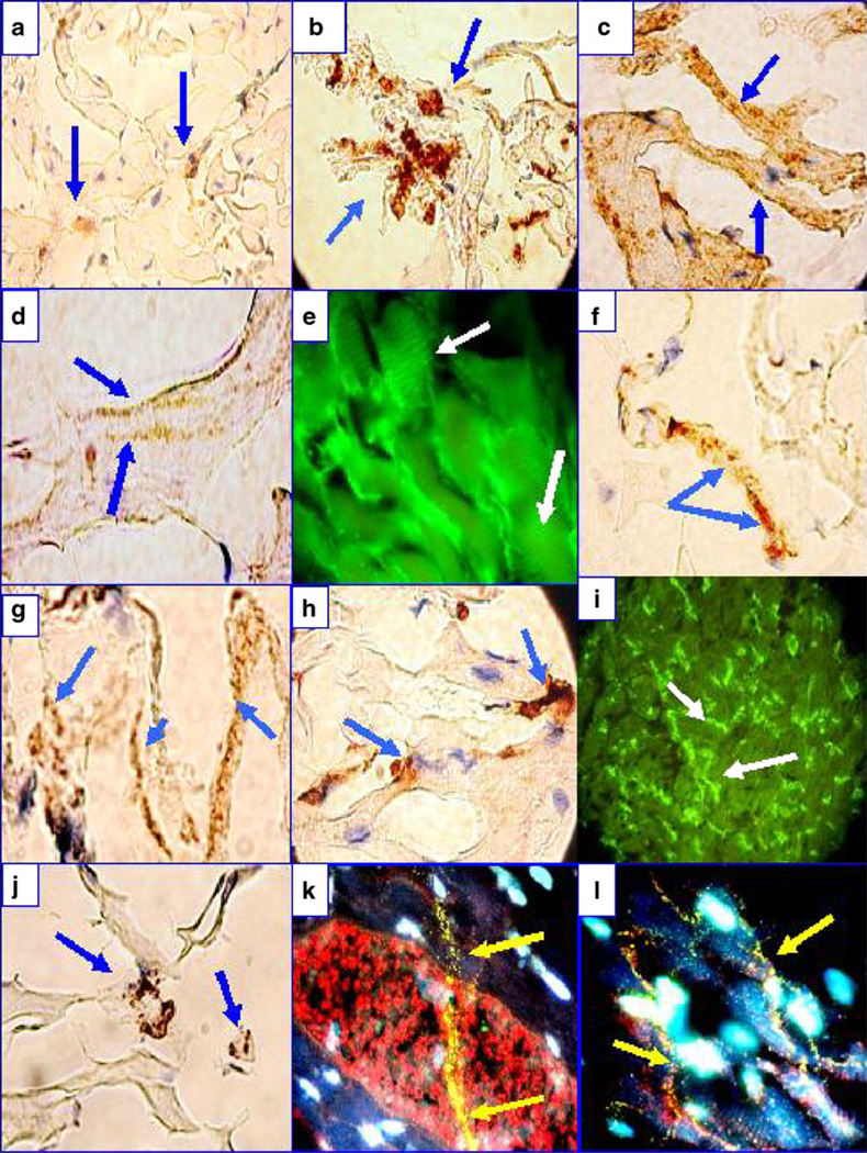
Fig. 3.
IHC and IIF of a heart from one El Bagre-EPF patient necropsy. a Anti-human IgG-positive IHC staining to the area composita of the heart (blue arrows; brown staining). b Positive IHC staining using anti-human HAM 56 antibody (brown staining; blue arrows). c Positive IHC staining against several areas of the heart using anti-human complement/C1q (blue arrows). d Positive IHC staining of the sarcomere using anti-human IgG (blue arrows). e IIF showing similar sarcomeric staining using anti-human IgG (white arrows). f Positive IHC staining of the heart using anti-human IgM (blue arrows). g Anti-human kappa light chains stained strongly positive (blue arrows). h Positive IHC staining for myeloperoxidase (brown staining; blue arrows). i DIF showing positive staining along intermyocyte junctions (white arrows). j Positive IHC staining for mast cell tryptase (brown staining; blue arrows). k IIF showing the area composita, highlighted in red using Texas red-conjugated anti-DP I–II and positivity to a long linear structure (yellow staining; yellow arrows), utilizing FITC-conjugated anti-human IgM antibody. The linear structure was also faintly positive with DP I and DP II antibodies, precisely colocalizing with El Bagre-EPF autoantibodies against the Purkinje fibers. The nuclei are counterstained in light blue with DAPI. l Same as k but at lower magnification

