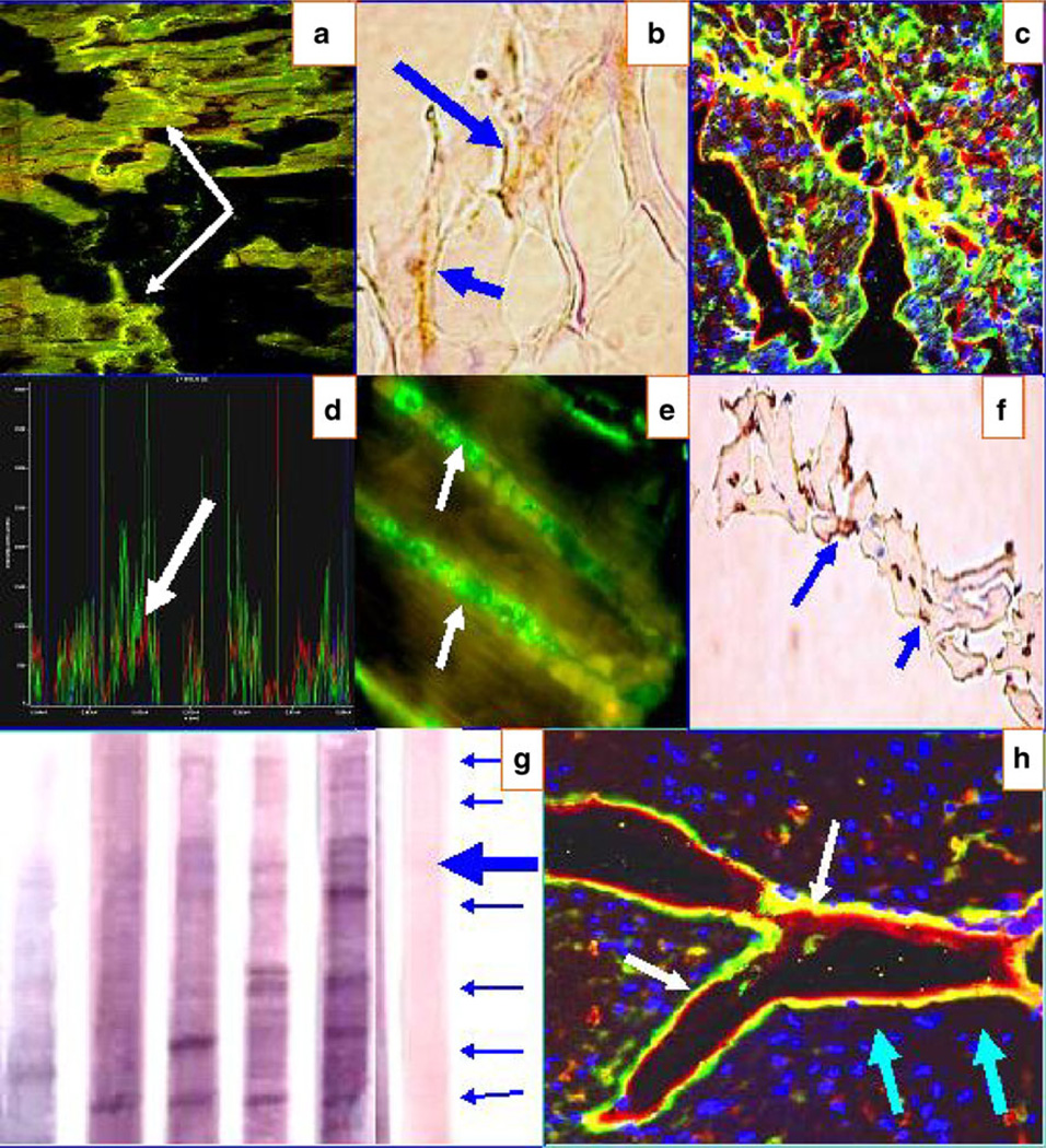
Fig. 4.
a CFM image showing positive staining to the Purkinje fibers (yellow bead-like dot staining) from one El Bagre-EPF patient serum (white arrows). b IHC from El Bagre-EPF patient necropsy heart staining positive to anti-human HLA DPDQDR antibody (brown staining; blue arrows). c Same as a at lower magnification. d CFM colocalization of the fluorescent peaks of the a/c data, showing that antibodies to Connexin 43 (red staining) colocalize precisely with the patient antibodies (green staining; white arrow). The blue peaks represent DAPI nuclear counterstaining. In e, we found positive reactivity to the lateral area of the myocytes (where mitochondria are located) using one El Bagre-EPF patient serum (white arrows). f IHC from El Bagre-EPF patient necropsy heart, displaying positive staining to anti-human HAM 56 (brown staining; blue arrows). g An IB of heart tissue extract. The blue arrows at the right show (in descending order) reactivity against 300, 250, 210, 190, 133, 120, and 97 kDa molecules in the sera from El Bagre-EPF patients. The large blue arrow highlights a triplet of positive antigens, aggregated in the 210-kDa area. One control from the endemic area also showed positive staining to an antibody of 250 kDa. h CFM image showing positive staining in heart tissue to ICAM-1/CD54 (red staining; light blue arrows). The yellow/green staining represents reactivity of the patient autoantibodies using FITC-conjugated anti-human IgG antibodies (white arrows). Nuclei are again counterstained with DAPI (dark blue staining)

