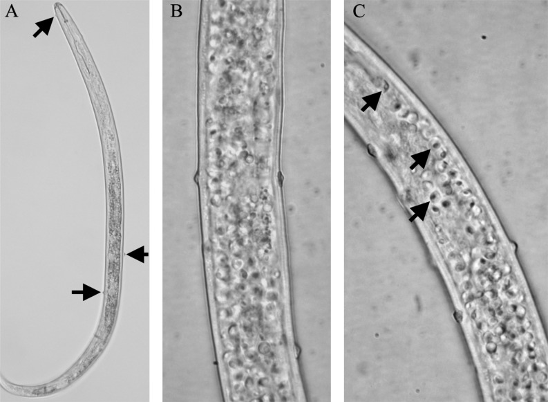
Fig. 1.
Brightfield micrographs of Pasteuria spp. Pr3 endospores attached to and infecting Rotylenchulus reniformis recovered from field soil in Huxford, AL. A) Arrows indicate endospores attached to the cuticle surface. B and C) Show endospores attached to the cuticle and the presence of mature endospores within the body cavity (indicated by arrows in C).

