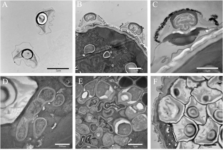
Fig. 6.
Transmission electron photomicrographs of Rotylenchulus reniformis Pasteuria Pr3 parasitizing its host. A) Free endospores. B-C) Pasteuria endospores bound to host cuticle in the first stage of infection. C) Shows development of the germ (penetration) tube at the base of the attached endospore indicating germination. D) Stage I of sporulation (mycelial stage) of development (center right) and a mature endospore (upper left) in the pseudocoelom of a single specimen. E) Stages I-V of sporulation. F) Fully mature Stage VII Pasteuria endospores.

