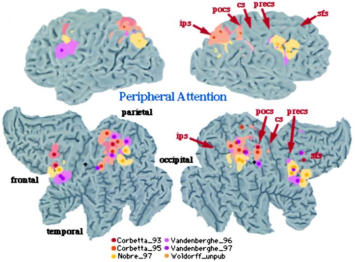
Figure 2.
3D rendering and 2D flattened surface of the Visible Man Brain, with the left hemisphere on the left. Lobes are indicated in 2D surface. Sulci are indicated as follows: sfs, superior frontal sulcus (s.); precs, precentral s.; cs, central s.; pocs, postcentral s.; ips, intraparietal s. Foci of activation during shifting attention [red (16), yellow (17), and orange (53)] and tonic attention [pink (24), violet (25), and light orange (Woldorff et al., unpublished data)].

