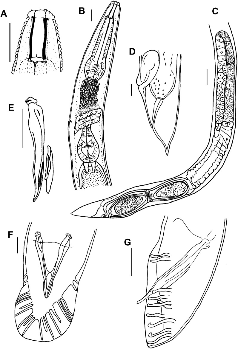
Fig. 1.
Parasitorhabditis frontali n. sp. Drawings with scale bars of 10 μm for A, B, D – F, and 20μm for C. A. Female stoma, lateral view, B. Female pharynx, lateral view, C. Female reproductive system, lateral view, D. Female tail, lateral view, E. Male spicule, slipper-shaped gubernaculum, lateral view, F. Male tail with spicules, ventral view, open bursal fan, fused spicule tips, 10 rays, 2 + (3 + 2) + 3 ray pattern. G. Male tail with spicules, lateral view, 10 rays, 2 + (4 + 1) + 3, with ray 6 trapped under ray 5 so that ray tip order is reversed from typical pattern.

