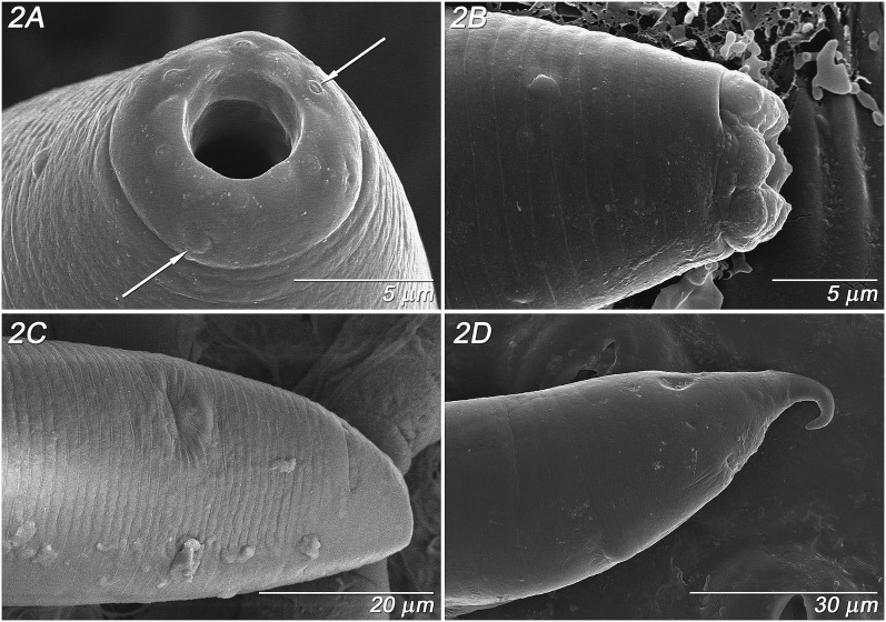
Fig. 2.
Parasitorhabditis frontali n. sp. LT-SEM images of female morphology. A. Pseudolips, face view, with cephalic papillae (left arrow) labial papillae (right arrow), B. Pseudolips, lateral view, C. Tail, ventro-lateral view with vulval slit (upper middle surface) and anal slit (upper right surface), D. Tail, lateral view with vulval slit (lower middle surface) and anal slit (upper right surface), curved tail spike.

