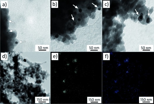
Figure 1.

Modification of C-120 type silica (specific surface area 114 m2 g−1) using triethoxysilane generates silicon hydride groups. These are subsequently used to reduce a silver nitrate solution to AgNPs, which are affixed to the top of silica. TEM images reveal the distribution of near-spherical AgNPs, appearing as darker contrast particles (white arrows) against the fused silica substrate, with particle sizes averaging a) 11 nm, b) 31 nm, and c) 45 nm. d) EDX mapping analysis reveals the corresponding location of silver and mercury in the TEM image for the e) Ag (Lα peak) and f) Hg (Mα peak) after their reaction and shows that the distribution of Hg correlates only to the location of AgNPs.
