
Fig. 1.
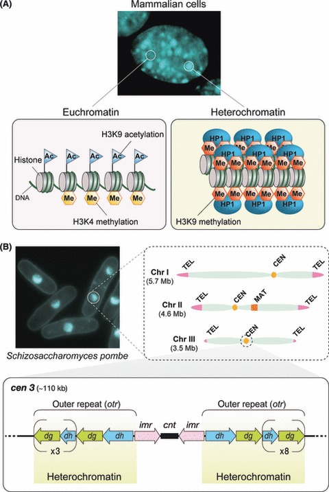
Heterochromatin and epigenetically silent regions of the genome. (A) Active protein coding genes are generally contained within euchromatin, typically modified by acetylation (Ac) of histone H3 lysine 9 (H3K9) and methylation (Me) of histone H3 lysine 4 (H3K4). In contrast, heterochromatin is generally considered to have a compact structure and can be observed as densely stained regions in nuclei. Heterochromatin is characterized by H3K9 methylation, which is recognized by Heterochromatin Protein 1 (HP1). The upper panel shows one complete 4′6′-diamidino-2-phenylindole dihydrochloride (DAPI)-stained nucleus (center) of a mouse NIH3T3 cell. (B) Schizosaccharomyces pombe chromosomes and heterochromatin. The S. pombe genome is organized across three chromosomes, with heterochromatin found at the telomeres (TEL), centromeres (CEN) and mating type region (MAT). Centromeric regions (lower panel) are arranged with a unique core centromere region (cnt and imr) flanked by heterochromatin covering the pericentromeric outer repeat regions (otr). The otr contains multiple copies of dg and dh repeat sequences that vary in number depending on the chromosome. The upper left panel shows S. pombe cells stained with Hoechst33342.